
Electron microscope
The electron microscope is a microscope that can magnify very small details with high resolving power due to the use of electrons rather than light to scatter off material, magnifying at levels up to 500,000 times.
History
The first electron microscope was built in 1931 by Ernst Ruska and Max Knoll at the Berlin Technische Hochschule. It was greatly developed through the 1950s and has allowed great advances in the natural sciences. The advantage of an electron beam is that it has a much smaller wavelength (see wave-particle duality), which allows a higher resolution - the measure of how close together two things can be before they are seen as one. Light microscopes allow a resolution of about 0.2 micrometer (200 nm, whereas electron microscopes can have resolutions as low as 0.1 nm (1 angstrom).
Process
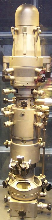
High voltage electron beams from a cathode are focused by magnetic lenses on to the specimen. They are then magnified by a series of magnetic lenses until they hit photographic plate or light sensitive sensors - which transfer the image to a computer screen. The image produced is called an electron micrograph (EM).
Types
The Transmission electron microscope (TEM) produces images by detecting electrons that are transmitted through the sample, while the Scanning electron microscope (SEM) produces images by detecting secondary electrons which are emitted from the surface due to excitation by the primary electron beam.
Generally, the TEM resolution is about an order of magnitude better than the SEM resolution, however, because the SEM image relies on surface processes rather than transmission it is able to image bulk samples and has a much greater depth of view, and so can produce images that are a good representation of the 3D structure of the sample.
A Scanning Transmission Electron Microscope (STEM) is a specific sort of TEM, where the electrons still pass through the specimen, but, as in SEM, the sample is scanned in a raster fashion.
Treatment
Samples viewed under an electron microscope may be treated in many ways:
- Cryofixation - freezing a specimen so rapidly, to liquid nitrogen temperatures, that the water forms vitreous (non-crystalline) ice. This preserves the specimen in a snapshot of its solution state. This technique produces the best specimen preservation, but isn't applicable to all specimens.
- Fixation - preserving the sample to make it more realistic. Glutaraldehyde - for hardening - and osmic acid - which stains lipids black - are used.
- Dehydration - the removing of water to be replaced with an embedding medium such as ethanol or propanone.
- Embedding - supports the tissue for sectioning in a resin such as araldite.
- Sectioning - produces thin slices for mounting. These can be cut on an ultramicrotome with a diamond knife to produce very thin slices.
- Staining - uses metals such as lead and uranium to reflect electrons to give contrast between different structures.
- Ion Beam Milling - thins samples until they are transparent to electrons by firing ions (typically argon) at the surface from an angle and sputtering material from the surface. A subclass of this is Focussed Ion Beam milling, where gallium ions are used to produce an electron transparent membrane in a specific region of the sample, for example through a device within a microprocessor.
Disadvantages
The
samples have to be viewed in vacuums, as the molecules that
make up air would scatter the electrons. This means that no living material can
be studied. Recent advances have allowed samples to be imaged using lower vacuums
and with partially hydrated samples, and the use of an electron transparent membrane
between a biological sample and the vacuum has been shown to allow fully hydrated
samples to be imaged, although there is a reduction in resolution and contrast,
and the intense radiation of the electron microscope is sufficient to cause cell
death.
The samples have to be prepared in many ways to give proper detail,
which may result in artifacts—objects that are purely the result of treatment.
This gives the problem of distinguishing artifacts from material, particularly
in biological samples.
There have been a few scientists, such as Dr. Harold Hillman, who believe that such artifacts are responsible for all the structures observed in biological samples by electron microscopy, rendering the techniques useless for these materials. Mainstream scientists maintain that the results from various preparation techniques have been compared, and as there is no reason that they should all produce similar artifacts, it is therefore reasonable to believe that electron microscopy features correlate with living cells. In addition, higher resolution work has been directly compared to results from X-ray crystallography, providing independent confirmation of the validity of this technique. Recent work performed on unfixated, vitrified specimens has also been performed, further confirming the validity of this technique.
Electron microscopes are also very expensive to buy and maintain.
See Category:Electron microscope images for images made with electron microscopes.